Plants are exposed to their environment in all types of situations. To survive and thrive, they have had to develop sophisticated solutions through thousands of years of evolution that will guarantee their lasting life, even during harsh conditions. Plants are natural smart factories of effective compounds with specific properties. One such marvel of nature is plant cell organelles called oleosomes, also known as oil bodies. While oleosomes are not new to the industry, the scientific knowledge base about them has progressed over the past 10+ years.
Log in to view the full article
Plants are exposed to their environment in all types of situations. To survive and thrive, they have had to develop sophisticated solutions through thousands of years of evolution that will guarantee their lasting life, even during harsh conditions. Plants are natural smart factories of effective compounds with specific properties. One such marvel of nature is plant cell organelles called oleosomes, also known as oil bodies. While oleosomes are not new to the industry, the scientific knowledge base about them has progressed over the past 10+ years.
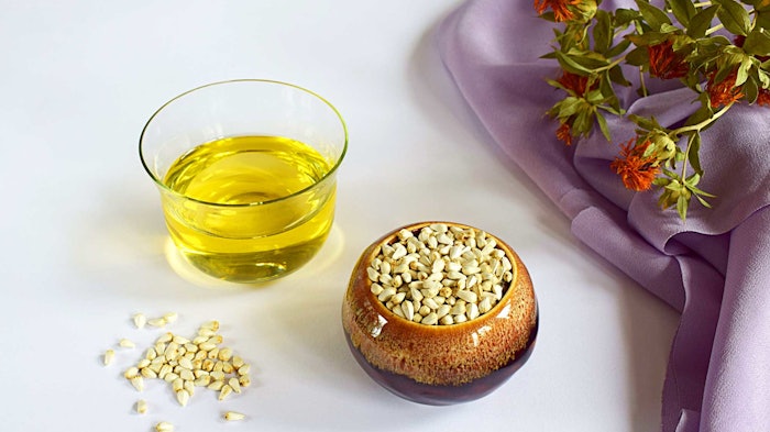
Safflower oleosomes are of interest in part because the plant seeds uniquely contain a large amount of oil bodies and low water content, resulting in decreased hydrolytic properties and lower protein degradation.1 The high oil content also creates low surface tension, increasing the oleosome’s ability to act as an emulsifier.1
As such, the present article explores safflower oleosomesa for their application in the personal care sector. Their structure and function are first described, followed by stability testing in emulsions. A clinical study using Confocal Raman Spectroscopy (CRS) also is performed to provide new insights into the hydrating and barrier protection potential of the ingredient; TEWL, skin water content and thickness, and ceramide and fatty acid measurements are described.
Plant Oleosomes 101
Oleosomes are natural oil droplets, abundant in plants and more specifically in seeds, with a typical diameter of 1-3 µm. The primary function of oleosomes is to safely store the seed energy source in the form of triacyglycerols (vegetable oil) during dormancy and use it during germination, and to protect the seeds against environmental stressors.
To achieve this, oleosomes are equipped with a sophisticated membrane that covers the triacylglycerols (TAGs) core, granting them physical and chemical stability. The membrane acts as a shield, preventing the oxidation of the TAGs. The membrane is dilatable and consists of a continuous monolayer of phospholipids that contains a number of proteins, mostly oleosins, embedded in it (see Figure 1).
The oleosome interfacial molecules create an interface around the oleosome lipid core that prevents their coalescence.2 The oil contributes up to 99% while the membrane constitutes only 1% of the total oleosome unit.3 TAGs, also known as triglycerides, are mainly constituted by a glycerol part in which each hydroxyl group is esterified to fatty acids.
Oleosins are amphiphilic in nature with long hydrophobic domains that are anchored into the oil phase, while their amphiphilic domains rest on the hydrophilic oleosome surface.4, 5 They play a major role in the stabilization and function of oleosomes.6 The inherent physical stability of oleosomes as provided by their interfacial molecules make them good candidates as bio-based alternative for oil and water emulsions.
Within the oil part, tocopherol or vitamin E occurs naturally. Tocopherol is a known antioxidant and its function is to prevent the rancidification of the unsaturated triglycerides stored within the oleosome.3
Safflower Oleosomes
For the present work, oleosomes were naturally and sustainably extracted from the seeds of safflower (Carthamus tinctorius). The herbaceous non-GMO plant is cultivated and harvested in California, USA, without the use pesticides and with low water requirements.
Extraction: A proprietary aqueous extraction cold process is implemented, comprising: grinding the seeds with aqueous medium, centrifugation and separating the liquids from the solids by centrifugation again before isolating the oleosomes. This process keeps the oleosomes fully intact, maintaining the micro-oil bodies as they occur naturally, with their full qualities and capabilities.
Furthermore, cold processing retains the antioxidants, vitamins and minerals. The result is a natural oil-in-water (o/w) emulsion combining naturality with natural high-performance properties.
Fatty acids and vitamin E: The resulting oleosomes contain tocopherol (vitamin E) and a major group of compounds: fatty acids (FAs), which are key components for skin health. The fatty acid fraction is dominated by the long-chain linoleic and oleic fatty acids (see Table 1). Oleic acid is monounsaturated whereas linoleic acid is polyunsaturated (PUFA).
Stable Emulsification
As noted, the safflower plant’s resilient oleosomes present unique properties for personal care and cosmetics: natural high emulsification capabilities plus vitamin E for skin health. The amphiphilic nature of the oleosin protein coat gives the oleosomes self-emulsifying properties for stable emulsions.3
Oleosomes can emulsify additional oil as well and rearrange around the newly created larger oleosome interface to form stable emulsions thanks to the dilatable membrane. When dispersed oleosomes in water are homogenized in the presence of free lipids, the oleosome membrane molecules redistribute, adjusting their interfacial molecular density on the new available interface and reaching a new equilibrium.7
Indeed, the described safflower oleosomesa can emulsify up to three times their weight in oil. Studies were performed reviewing the stability of the simplest emulsions using oleosomes and oils at different polarity levels and different ratios (see Table 2). The oils included: Paraffinum liquidum, decyl oleate, caprylic/capric triglyceride, safflower oil and C12-15 alkyl benzoate).The stability was evaluated by two methods: 1) centrifugation: 3,500 rpm for 30 min, Turbiscan analysis, coloration, microscope observation; and 2) after 30 days at 40°C in an incubator; Figures 2a-b and 3 summarize the results.
The addition of a polymer or rheological modifier enabled stable emulsions with a ratio of oleosome:oil of 1:3. The safflower oleosomes were able to replace emulsifier systems within the range of 5 < HLB < 15. Furthermore, the obtained bio-based emulsions offered convenience in that they required no additional emulsifiers or heat during the homogenization step.
Barrier Protection Efficacy
The primary function of skin surface lipids is to form a natural protective barrier8 and FAs play an important role. The hydrophilic groups of long-chain FAs in particular, such as oleic, linoleic, palmitic and others, combine with moisture in skin and retain it, lubricating skin while reducing TEWL.9
Humans can synthesize monounsaturated FAs, which are mainly of a long-chain structure in skin, but they cannot produce PUFAs such as linoleic essential FA (EFA); they must be supplemented. These intercellular lipids, together with stratum corneum cells, constitute the well-known “brick-and-mortar” structure of skin; the bricks being flattened, anucleated corneocytes and the mortar being the extracellular multilamellar lipid-enriched matrix.10, 11
TEWL: Considering the FA-rich content of safflower oleosomes (see Table 1), a clinical study was conducted in 20 volunteers to compare the effects of the oleosomes versus commonly used petroleum-derived ingredients on water loss from skin. Four basic test formulas (see Formula 1) containing the safflower oleosomes, petrolatum, mineral oil or a 50/50 mineral oil/petrolatum blend were applied.
On day one, the initial TEWL values for each subject were recorded, then 0.30 g of each product was dispensed onto randomized 4 cm x 4 cm test sites (two per volar forearm with the center site on each arm serving as the untreated control). The products were rubbed into the skin using a finger cot.
Twenty-four hours later, the TEWL measurements were made on all test sites. The products were then re-applied and the measurements made 24 hr later. The test ended after 16 days.
The percent reduction in water loss was determined by comparing the TEWL values for the two untreated controls to the TEWL values of the four test sites. TEWL values for each test product were divided by the TEWL values of the average of the two controls and subtracted from 100 to yield a percent TEWL reduction between the test product and the untreated control. Low TEWL values for any test product (indicating a strong occlusive barrier) would thus be expressed as a high percent reduction in TEWL.
TEWL was measured using confocal Raman spectroscopy (CRS) (see Figure 4). CRS is a non-invasive tool and the most frequently used Raman technique to investigate the penetration of exogenous substances into skin or to analyze the skin in a healthy or diseased state;12 it also can directly and accurately measure water molecules in the skin. In this case, CRS indicated the formulation containing the oleosomes showed the greatest reduction in TEWL especially in the early days of the trial; up to a 40% reduction at day two.
Skin hydration: An additional clinical study sought to determine if the safflower oleosomes could achieve ceramide-like effects since these lipid components of the skin barrier enhance its function. Thus, the effects of the oleosomes on water content in skin were compared with those of ceramide NP. Two test formulas (see Formula 2) were developed and applied by 10 female volunteers (ages 37 to 53) over the course of 28 days. Figures 5a-b represent the averaged water content (µg) per cm2 of stratum corneum (SC) before (day 0) and after 28 days of product application.
After 28 days, the safflower oleosome formula significantly (p < 0.05) increased the hydration (water content) of skin by 21.5%, compared with the ceramide formula, which increased it by 18.8%. More specifically, the water content increased 147.7 µg per cm2 of skin with the oleosomes versus 124.9 µg per cm2 with the ceramide, marking a significant increase.
SC thickness: CRS was additionally used to measure the thickness of the SC in vivo after treatment with the described safflower oleosome or ceramide formulas. Figures 6a-b show the averaged SC thickness before (Day 0) and after 28 days of test product application. After 28 days of test product application, the safflower oleosomes increased the SC thickness by 18.1%, whereas the ceramide formula increased it 21.5%; more specifically, the SC thickness increased an average of 2.8 µm with the oleosomes and 3.2 µm with the ceramide. These results verified the ceramide-like effects of the oleosomes.
Ceramide and fatty acid content: Lastly, CRS again was used to directly measure the ceramide and fatty acid content levels in the stratum corneum before/after treatment with the test formulas. Figure 7 shows the average amount of ceramides and fatty acids (arbitrary units) per cm2 of SC before (Day 0) and after 28 days of applying the safflower oleosome or ceramide formula. Both formulas increased ceramide and fatty acid levels in the stratum corneum, with a higher increase (+4.9%) observed for the safflower oleosomes: oleosomes increased FA content in the SC by 10% (p < 0.07) while ceramide NP results were not significant (p = 0.45).
Discussion
Formulas containing the test ingredient contain tens of billions of oleosomes per gram of ingredient. Upon application, these would collapse, disrupting their membranes and releasing moisturizing oils and vitamin E onto the skin to deliver skin barrier benefits.
Thus, the oleosomes would impart restorative action on the lipid structures of the stratum corneum due to their structure as a whole. Safflower oil, the main component of oleosomes, is a triglyceride, rich in lipid fractions, even unsaturated ones, that play a fundamental role as precursors in the skin ceramide synthesis.
Conclusion
While oleosomes are not novel to the cosmetic industry, the knowledge base about them has advanced over the past decade or more. Within this context, safflower oleosomes were explored as described here for their unique structure and potential in personal care applications.
Their self-emulsifying/emulsifying properties were shown to support the creation of stable o/w emulsions, incorporating up to three times their weight in external oils. They also delivered fatty acids to the stratum corneum, reducing TEWL and increasing the average water content, skin thickness and FA/ceramide content in skin, as demonstrated in vivo through CRS.
Safflower oleosomes therefore combine naturality and sustainability with high performance for multifunctional benefits in modern formulations.
a Hydresia SF2 (INCI: Carthamus Tinctorius Oleosomes (Safflower) (and) Water (Aqua) is a product of Sharon Personal Care.
References
1. Patel, P., Wang, J.Y., Mineroff, J., et al. (2024). The potential cutaneous benefits of Carthamus tinctorius oleosomes. Arch Dermatol Res, 316, 26.
2. Ntone, E., Yang, J., Meinders, M.B.J., Bitter, J.H., Sagis L.M.C. and, Nikiforidis, C.V. (2023). The emulsifying ability of oleosomes and their interfacial molecules. Colloids and Surfaces B: Biointerfaces, vol 229 113476.
3. Palak, A., Yadav, K.S., Rai, V.K. (2021). Natural and multifunctional colloidal carriers: A new prospective in drug delivery. Pharmaspire, 13(4) 168-176.
4. Nikiforidis, C.V. (2019). Structure and functions of oleosomes (oil bodies). Adv Colloid and Interface Sci, vol. 274 102039.
5. Karefyllakis, D., van der Goota, A.J. and Nikiforidis, C.V. (2019). The behavior of sunflower oleosomes at the interfaces. Soft Matter, 15 4639.
6. Maurer, S., et al. (2013). The role of intact oleosin for stabilization and function of oleosomes. J Phys Chem B, 117 44, 13872 - 13883.
7. Ntone, E., et al. (2023). The dilatable membrane of oleosomes allows their in vitro resizing and triggered of lipids. Soft Matter, 19, 6355 - 6367.
8. Proksch, E., Brandner, J.M. and Jensen, J.M. (2008 Dec). The skin: An indispensable barrier. Exp Dermatol, 17(12) 1063-72.
9. Yang, M., Zhou, M. and Song, L. (2020). A review of fatty acids influencing skin condition. J Cosmet Dermatol, 19 3199 - 3204.
10. Jungersted, J.M., Hellgren, L.I., Jemec, G.B. and Agner, T. (2008 May). Lipids and skin barrier function — A clinical perspective. Contact Dermatitis, 58(5) 255 - 62.
11. Lefèvre-Utile, A., Braun, C., Haftek, M. and Aubin, F. (2021, Oct 28). Five functional aspects of the epidermal barrier. Int J Mol Sci, 22(21).
12. Lunter, D., Klang, V., Kocsis, D., Varga-Medveczky, Z., Berkó, S. and Erdő, F. (2022 Sep). Novel aspects of Raman spectroscopy in skin research. Exp Dermatol, 31(9) 1311 - 1329.


